The activity shown in the cartoon is actually a montage, made up of recordings from different episodes of the motor programme. The FETi and FlTi activities were recorded simultaneously from one episode, but the FI and M activities were recorded separately from different animals. This is because of the technical difficult of recording from several neurons at once.
The recordings are made from grasshoppers performing hind-leg kicks, rather than grasshoppers actually jumping. This is for the obvious reason that it is impossible to keep microelectrodes in the animal if it jumps freely. However, there is good evidence that the motor programme driving the hind leg extension is the same in jumping and kicking (and, surprisingly, swimming).
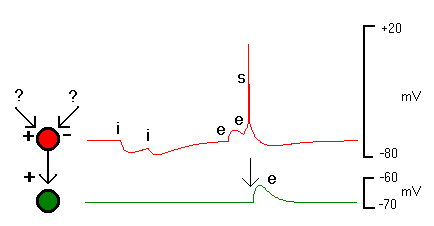
The recordings in the cartoon show the membrane potentials of the 4 neurons - i.e. the voltage difference between the inside and the outside of the cells, plotted against time. When they are not doing anything, neurons have a resting potential of about -70 mV (the inside is negative relative to the outside). Synaptic potentials arrive as a result of input connexions from other neurons, and briefly shift the membrane potential away from its resting state. If the synapse is excitatory, the membrane potential tends to shifts positive. If the synapse is inhibitory, the membrane potential tends to shift negative.
A neuron may receive synaptic input from many other neurons. It integrates the excitatory and inhibitory inputs, and if the resulting change in membrane membrane potential goes sufficiently positive (above threshold), the neuron itself produces a nerve impulse, or action potential (commonly called a spike). The action potential, unlike the synaptic potentials, travels down the axon of the neuron, and either causes a synaptic input to another neuron (if the spiking neuron is an interneuron), or a muscle contraction (if the spiking neuron is a motorneuron).
In the picture above the red interneuron neuron gets inhibitory (i) and excitatory (e) synaptic inputs from unknown (?) sources. The two excitatory inputs occur close together in time and add together (temporal summation), causing the red neuron to spike (s). The red neuron makes an excitatory synaptic connexion to the green neuron.
The key point is that neurons integrates the synaptic input they receive, and only produce output themselves if the summed input is above their spike threshold.
The traces in the cartoon show intracellular recording made from the cell bodies of the motorneurons, and the neuropilar segment of M. The spikes are attenuated in amplitude, because the recordings are made from regions in the neuron which do not produce full action potentials. In each record the maximum vertical excursion is about 30 mV.